COBRE
Department of Ophthalmology
University of Oklahoma Health Sciences Center
HOME | PI | PJIs | CORES | MENTORS | IAC | EAC | SPOTLIGHT | SEMINARS | CALENDAR | AFFILIATES
John Ash, Ph.D.
Wei Cao, Ph.D.
Michael Ihnat, Ph.D.
Yun Le, Ph.D.
Raju Rajala, Ph.D.
PROMISING JUNIOR INVESTIGATOR
JOHN ASH, PH.D.
COBRE Research Project
JOHN ASH, PH.D.
COBRE Research Project
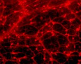
Regulation of Vascular Development in the Mouse Retina.
(Mentor, Hiroyuki Matsumoto, PhD)
Neovascularization of the choroid leads to the majority of catastrophic vision loss in age-related macular degeneration, the most common cause of severe vision loss in the United States. Retinal neoangiogenesis is the cardinal and common histopathologic and clinical hallmark of eye diseases that include sickle cell retinopathy, diabetic retinopathy, branch vein occlusion, and retinopathy of prematurity. Diabetic retinopathy is the most common cause of severe vision loss in working age Americans and is unusually prevalent in states with large Native American populations such as Oklahoma. These diseases all are characterized by the loss of normal ocular blood vessels and the subsequent uncontrolled growth of abnormal blood vessels in the retina, subretinal space, and/or vitreous of the eye. In contrast to humans, mice can replace lost or damaged retinal vessels and can spontaneously resolve blood vessels in the vitreous. We hypothesize that the rodent retina is competent to promote retinal vascular regrowth through the expression of genes that are essential regulators and effectors of blood vessel growth and stability. To better understand the endogenous mechanisms for long-term protection from proliferative retinopathy in mice, our goal is to identify the molecular regulators and effectors of vascular growth in the mouse retina. In the current proposal, we will use transgenic mice in which we can induce the expression of TGF- 1 (aim 1), or LIF (aim 2) in retinal photoreceptors, so that we can turn off retinal competence for vascular development at ages of our choosing (aims 1 and 2). With these mice, we will test the hypothesis that cytokines reduce growth of vascular endothelial cells or increase vascular degeneration. We will also test the hypothesis that cytokines down-regulate the expression of specific genes that are necessary for retinal vascularization (aim 3). Dr. Hiro Matsumoto will mentor this project. Dr. Ash has extensive training in generating and characterizing transgenic mice expressing different transgenes in the lens. It is anticipated that the goals of this proposal are feasible based on this experience; however, the project will benefit greatly from the research experience of Dr. Matsumoto. He has twenty years experience in analyzing vision-related gene expression, and is the director of both the P30 biochemistry module, and the mass-spec/proteomic facility at OUHSC. He is an ideal mentor, as Dr. Ash serves as collaborator on Dr. Matsumoto's project involving proteomic analysis of genes expressed in rodent retinal development. This collaboration will facilitate the goals of this proposal. Dr. Matsumoto will provide the expertise for the proteomic analysis outlined in this proposal. The experiments in this proposal will require several of the supported core modules. The Microinjection Core module will be used to generate the new transgenic mouse models, which will be housed in the Animal Core module in the DMEI vivarium. He will heavily use the Image Acquisition and Processing Core module to image, and quantify changes in retinal vascular development. In addition, the Proteomics/Bioinformatics and Microarray Core modules will be heavily used for gene expression analysis. Dr. Ash will make moderate use of the Molecular Biology Core module, which will be used to cryopreserve existing and new transgenic models.
Please send comments,
questions, or error reports to Holly-Whiteside@ouhsc.edu

Copyright © 2003 The Board of Regents of the University of Oklahoma, All Rights Reserved
University of Oklahoma Disclaimers
Copyright © 2003 The Board of Regents of the University of Oklahoma, All Rights Reserved
University of Oklahoma Disclaimers