COBRE
Department of Ophthalmology
University of Oklahoma Health Sciences Center
HOME | PI | PJIs | CORES | MENTORS | IAC | EAC | SPOTLIGHT | SEMINARS | CALENDAR | AFFILIATES
Molecular Biology
Image Acquisition and Production
Animal Core
Lipid Analysis
IMAGE ACQUISITION AND PRODUCTION MODULE
The purpose of this module is to provide quality state of the art instrumentation and expertise to obtain microscopic images with light, fluorescent and Nomarski optics; to archive the images in a retrievable format, to provide for morphometric analysis and to produce publication quality prints. The Module comprises 600sq ft in the basement of the Dean McGee Eye Institute. The Module equipment consists of optical image acquisition devices interfaced with computer systems for image storage and manipulation. In addition, the facility contains paraffin embedding equipment, microtomes, a cryostat, a refrigerated chromatography cabinet, a thermal cycler, a tissue processing hood, a refrigerated intermediate speed centrifuge, protein and DNA electrophoresis equipment, electro-blotting equipment, water baths and an incubator. In an adjacent, "clinical pathology room", we have access to an automated tissue processing station, two additional paraffin microtomes, an automated histochemical (H&E) slide stainer and an automated immuno-cytochemical staining system.
Module director: James F. McGinnis, PhD is the overall Director of the module. Dr. McGinnis is currently director of the microscopy facility at the DMEI and has extensive experience in: fixation, embedding and sectioning of ocular tissues, photomicroscopy, immunocytochemistry, generating primary cell cultures of neuronal tissue, in situ hybridization and a variety of techniques in molecular biology. He will oversee the image acquisition module, the Systems Manager, the histology technicians and coordinate the assignment and scheduling of projects from the core participants and the PJIs through the systems manager. The current histology technician is working full time on the projects of the PIs who are members of the Core and the TBA Histology Technician will provide histological services for the PJIs.
| Services Provided Samples will either be fixed by the Histology Technician or if more appropriate (e.g. timed tissue culture experiment or fresh surgical material) will be fixed by the investigator. Using automated systems, the tissue will be embedded in paraffin, sectioned and mounted on slides. Routine histochemical staining will be performed to assess the morphological quality of the sections. For immunocyto-chemistry, the investigator will provide any primary antibodies and the Histology Tech will provide any secondary antibodies and/or linkers (e.g. streptavidin) necessary for visualization by light or fluorescence microscopy. For in situ hybridizations, the investigator will provide the nucleic acid probe to be used and information concerning the conditions for hybridization, if known. The histology tech will actually perform the hybridizations and subsequent visualization reactions. |
 |
Sections will be viewed under the microscope, initially by the Systems Manager and the Histology Tech. The investigator will be shown some representative fields for his/her determination as to the experimental value of the sections. Based on the investigator's selection, preliminary images will be acquired using the microscope, put into the computer and catalogued. If unique knowledge is required to select fields or if a significant number of images are to be acquired, the investigator will be taught to use the microscope, camera, scanner, printer etc. If appropriate, the images can be processed or manipulated (arrows, labels, legends, etc.) and preliminary pictures printed out on an Epson Stylus color printer ($0.20/ page). The images for publication quality prints ($2.80/page) will be selected by the investigator and printed on the Kodak dye sublimation printer. The images will be emailed to the investigator or given on a Zip disk or a compact disc and he/she will be able to process them on one of the workstations using Licenced Copies of Metamorph software or on any computer using Photoshop or some other print management software. A copy of all original images will be stored and catalogued on the internet server. If the files become to large for the server, the investigators will be asked to "cull" them or purchase an additional storage device.
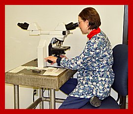 |
Equipment in the Image Core Module equipment is state of the art quality. This scope has unique optical benefits including the highest numerical apertures, the longest working distances, the best chromatic corrections available and the highest transmission UV optics. It is completely equipped for all light microscopic techniques such as Brightfield, Phase, Fluorescence, DIC, and APO Nomarski. In addition, it is equipped with filter cubes to enable double and triple fluorescence labeling to be directly viewed and photographed. |
The Princeton Instruments' Digital MicroMAX is a highspeed, low-noise CCD camera system designed with "Roper" technology for acquisition of low light fluorescent imaging applications and all light microscopic techniques. Because of its compact size, the MicroMAX camera attaches directly to the microscope with a cooled CCD, advanced exposure-control timing, video output, and sophisticated readout capabilities, making it ideal for high resolution images. It has low noise / high sensitivity, sensor-specific cooling, high-QE CCD sensors and dual-digitizer capability. This is the best overall digital camera on the market today. Its cooled color digital camera provides brilliant low light level color images especially for high magnification fluorescence applications The software allows the user to choose between shorter exposure, lower quality images and longer exposure high quality low light level images. The CCD chip has a resolution of 1315 x 1035 pixels which provides a much higher image capture resolution than that achieved by an analog camera-frame grabber combination. This camera is attached to the Nikon Eclipse 800 light microscope.
An Argus II scanner is also attached to the computer. This scanner can acquire transparent images from any photographic negative or print, including electron microscope, slit-lamp or fundus photograph, etc, at a resolution of 1024 by 1024. In addition, an image of an entire whole mount sample on a 1" x 3" microscope slide can be acquired using the Polaroid SprintScan 35 slide scanner and PathScan Enabler resulting in a high resolution 2700dpi graphic image which can be stored, catalogued or manipulated using the "Electro Windows Professional Digital Image Management and Archiving Software". This software package enables up to 15,000 images to be stored per catalogue with an unlimited number of catalogues (dependent on the size of the hard disk or server) and no longer than 5 seconds to search any catalogue for a specific image. Any image can then be E-mailed or printed on the Kodak XLS 8650 PS dye sublimation printer. This printer produces publication quality 81/2 x 11 inch photographs from digital image files in 70 seconds.
The computer is equipped with a Pentium III chip, operated at 800 MgHerz with a 1600 x 1280, 24 bit color display card which projects an image to a 21 inch (.25" dot pitch) Viewsonic color monitor; a 40 gigabyte hard drive, a Zip drive, a read/write CD recorder and a direct connection to the internet. This enables any of the users to download their images to 100 MB discs, 650 MB cds or directly onto their computers through the internet. The images can then be modified, labeled, made into collages and/or catalogued on the users computer and brought back for final printout on the Dye Sublimation printer.
The MetaMorph Imaging System Software provides extensive analytical functions to measure areas and distances, count objects or cells, and measure or graph intensity and optical densities. Spatial parameters like area, distance, diameter, perimeter, object counts, and many other variables are measured and logged via two basic methods: 1) interactive region measurements where the user creates line or area regions and MetaMorph displays spatial or intensity data based on those regions, or 2) automated multiple object measurements utilizing an intensity threshold. This system is licenced on each of the three work stations.
Work stations: There are a total of three workstations, including the one connected directly to the microscope. Each is similarly equipped with hardware and software including the sophisticated image analysis program "Metamorph". The second workstation is set up within the Image Core space but outside of the microscope room while the third is located on the fourth floor. All are connected to the internet. These independent workstations allow microscopic images to be viewed, organized, and analyzed without restricting the use of the microscope by others.
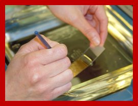
Tissue preparation: The core has a fully automated tissue processing station which allows fixation, dehydration, clearing, infiltration with paraffin over a 12 - 24 hour period to ensure complete reproducibility of the preparation step. Tissues are oriented and embedded in paraffin blocks using a Leica paraffin embedding center. This system is operated by the histology technician but is easy to use and available for direct use by others on a first-come, first-serve basis with a sign-up sheet when necessary.
Paraffin sections: (0.5-60 microns) can be cut on a fully automated microtome (Reichert 5540) or on the three other manual paraffin microtomes. These instruments are available to trained users a sign up basis or are used by the histology technicians to generate sections.
Cryostat sections: are prepared using a "Leica 1800 Cryocut" cryostat which allows users to obtain frozen (2-60 micron) sections of fixed or unfixed tissues. Sample preparation includes freezing and embedding the tissue in freezing medium. Trained users or the Histo tech operate the cryostat.
Histochemical staining: The facility includes a completely automated histochemical staining system (Ventana) which can take 350 slides simultaneously through the deparaffination, rehydrating and staining with H&E or Toluidine blue. This piece of equipment is also available to trained users on a signup basis or is used by the Histo tech.
Immunoctyochemical staining: The core has an automated Ventana immunocyto-chemical staining system which is available to the users of the Image and acquisition core facilities. This unit is operated by the histo tech and is set up for handling large numbers of slides stained with the same antibody at any one time. Manuals systems are also available with the user providing the primary antibody whereas all other reagents will be provided by the core.
Please send comments,
questions, or error reports to Holly-Whiteside@ouhsc.edu

Copyright © 2003-2012 The Board of Regents of the University of Oklahoma, All Rights Reserved
University of Oklahoma Disclaimers
Copyright © 2003-2012 The Board of Regents of the University of Oklahoma, All Rights Reserved
University of Oklahoma Disclaimers